Imagine trying to breathe while your lungs are wrapped in a tight, fluid-filled sack. That is exactly what happens with pleural effusion, a condition where excess fluid builds up in the pleural space-the thin gap between the lung and the chest wall. It’s not just an uncomfortable side effect; it’s a serious medical signal that something else is wrong inside your body. Whether caused by heart failure, cancer, or infection, this buildup restricts lung expansion, leading to shortness of breath, sharp chest pain, and a dry cough. Understanding why it happens, how doctors drain it safely, and most importantly, how to stop it from coming back, can be the difference between a manageable chronic condition and a life-threatening emergency.
The Two Main Types: Transudate vs. Exudate
To treat pleural effusion effectively, you first need to know what kind of fluid is sitting in your chest cavity. Doctors don’t just guess; they classify the fluid into two distinct categories based on its chemical makeup. This distinction dictates every step of your treatment plan.
Transudative effusions occur when systemic forces push fluid out of blood vessels and into the pleural space. Think of it like a leaky pipe due to high pressure elsewhere in the system. The most common culprit here is congestive heart failure, which accounts for nearly 90% of these cases. When the heart struggles to pump efficiently, pressure backs up into the lungs, forcing fluid out. Other causes include liver cirrhosis (where low protein levels fail to keep fluid in the veins) and nephrotic syndrome (kidney disease causing protein loss). In these cases, the fluid itself isn’t infected or inflamed; it’s just water displaced by pressure imbalances.
Exudative effusions, on the other hand, result from local inflammation or injury directly affecting the pleura. Here, the capillaries become "leaky" because of damage or irritation. Pneumonia is the leading cause, responsible for 40-50% of exudative cases. Malignancy (cancer) follows closely, accounting for 25-30%. Other triggers include pulmonary embolism, tuberculosis, and autoimmune diseases like rheumatoid arthritis. Because this fluid contains higher levels of proteins, cells, and enzymes, it’s thicker and more complex to manage than transudate.
| Feature | Transudative Effusion | Exudative Effusion |
|---|---|---|
| Primary Cause | Systemic pressure changes (e.g., Heart Failure) | Local inflammation or malignancy (e.g., Pneumonia, Cancer) |
| Fluid Appearance | Clear, straw-colored, watery | Turbid, bloody, or purulent |
| Protein Content | Low (Ratio < 0.5) | High (Ratio > 0.5) |
| LDH Levels | Low (Ratio < 0.6) | High (Ratio > 0.6 or > 2/3 upper limit normal) |
| Treatment Focus | Diuretics, managing underlying organ function | Antibiotics, chemotherapy, drainage procedures |
Distinguishing between these two relies on Light's Criteria, a diagnostic standard established in 1972. If any one of three specific ratios in the pleural fluid exceeds a certain threshold, the effusion is classified as exudative. This test has a sensitivity of 99.5%, making it the gold standard for diagnosis. Missing this distinction can lead to treating the symptoms while ignoring a deadly underlying cause, such as a hidden tumor.
Thoracentesis: How Fluid Is Removed Safely
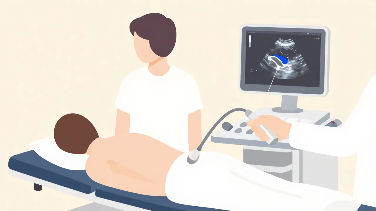
When the fluid buildup becomes severe enough to cause breathing difficulties, or when the doctor needs a sample to determine the cause, a procedure called thoracentesis is performed. This involves inserting a needle or catheter through the chest wall into the pleural space to drain the fluid. While it sounds invasive, it is a routine and highly effective intervention when done correctly.
The critical factor in modern thoracentesis is ultrasound guidance. Gone are the days of blind needle insertions. Ultrasound allows the physician to visualize the exact depth and location of the fluid pocket, avoiding vital structures like the liver, spleen, or intercostal arteries. Studies show that using ultrasound reduces complication rates dramatically-from 18.9% down to just 4.1%. Without it, the risk of accidental puncture increases significantly.
During the procedure, the patient usually sits upright and leans forward. The skin is numbed with local anesthesia. An 18-22 gauge needle is inserted into the mid-axillary line (the side of the chest), typically between the 5th and 7th ribs. For diagnostic purposes, only 50-100 mL of fluid is needed. However, if the goal is symptom relief, doctors may remove up to 1,500 mL in a single session. Removing too much fluid too quickly can cause re-expansion pulmonary edema, a rare but serious condition where the lung swells with fluid after being compressed for too long. To prevent this, physicians monitor the patient closely and may stop drainage if the patient feels sudden chest tightness or coughing.
Complications do happen, though they are less common now. The most frequent issue is a pneumothorax (collapsed lung), occurring in 6-30% of cases without ultrasound, but dropping significantly with imaging guidance. Bleeding and infection are also risks, albeit lower (1-2%). After the procedure, the site is bandaged, and a chest X-ray is often taken to ensure the lung has re-expanded properly and no air has leaked into the space.

Stopping the Cycle: Prevention of Recurrence
Draining the fluid provides immediate relief, but it does not cure the underlying problem. In fact, if the root cause isn’t addressed, the fluid will return-often within days. Preventing recurrence requires a strategy tailored specifically to the type of effusion.
For malignant pleural effusions (fluid caused by cancer), recurrence is extremely high. Without further intervention, 50% of patients see the fluid return within 30 days of thoracentesis alone. To stop this, doctors use two main approaches:
- Pleurodesis: This procedure aims to fuse the lung to the chest wall, eliminating the space where fluid can collect. A sclerosing agent, most commonly sterile talc, is injected into the pleural space. This causes inflammation that scars the layers together. Talc pleurodesis has a success rate of 70-90%. However, it can be painful and requires a hospital stay of several days while a chest tube remains in place.
- Indwelling Pleural Catheters (IPC): These are small, flexible tubes implanted under the skin that allow patients or caregivers to drain fluid at home. Recent data suggests IPCs are superior for many patients, offering an 85-90% success rate in controlling symptoms over six months. They reduce hospital stays from an average of 7.2 days to just 2.1 days, improving quality of life significantly for those with advanced cancer.
For heart failure-related effusions, the focus is entirely on medical management rather than surgical intervention. Optimizing medication regimens-including diuretics (water pills), ACE inhibitors, and beta-blockers-is key. When therapy is guided by biomarkers like NT-pro-BNP, recurrence rates drop from 40% to under 15% within three months. In these cases, repeated thoracentesis is rarely necessary unless the heart failure is acute and unresponsive to drugs.
Parapneumonic effusions (those caused by pneumonia) require aggressive antibiotic treatment. If the fluid becomes complicated (indicated by a pH level below 7.20 or glucose below 60 mg/dL), simple antibiotics aren’t enough. Drainage via a chest tube is mandatory to prevent empyema-a collection of pus in the pleural space-which occurs in 30-40% of untreated complicated cases. Early drainage saves lives and prevents long-term lung scarring.
When to Seek Immediate Help
Pleural effusion symptoms can escalate quickly. You should seek emergency care if you experience:
- Sudden, severe shortness of breath that doesn’t improve with rest.
- Sharp, stabbing chest pain that worsens when you take a deep breath or cough.
- Fever combined with chills and a productive cough (suggesting infection).
- Blue tint to lips or fingernails (cyanosis), indicating low oxygen levels.
Even if you have a known history of heart failure or cancer, new or worsening breathlessness warrants an immediate call to your healthcare provider. Ignoring these signs can lead to respiratory failure or sepsis, depending on the underlying cause.
Living with Pleural Effusion: Long-Term Outlook
The prognosis for pleural effusion varies wildly depending on the etiology. For patients with heart failure, managing the effusion is part of broader cardiac care, and survival rates remain tied to heart health. For those with malignant effusions, the outlook has improved slightly over the last decade, with 5-year survival rates rising from 10% to 25% thanks to better targeted therapies. However, untreated malignant effusions can reduce median survival to just four months, highlighting the importance of timely intervention.
Advancements in personalized medicine mean treatments are becoming more precise. For instance, pleural manometry (measuring pressure during drainage) helps predict complications before they happen. Additionally, the shift toward outpatient management with indwelling catheters empowers patients to maintain daily activities without frequent hospital visits. The goal is no longer just draining fluid-it’s preserving lung function and quality of life for as long as possible.
How long does it take to recover from a thoracentesis?
Most patients feel relief from breathing difficulties immediately after the procedure. Physical recovery is quick; you can usually go home the same day. However, you should avoid heavy lifting or strenuous activity for 24-48 hours to prevent bleeding at the insertion site. Full healing of the puncture wound takes about a week. If you experience persistent pain, fever, or increased shortness of breath, contact your doctor immediately.
Can pleural effusion go away on its own?
Small, asymptomatic effusions related to mild heart failure or minor infections may resolve on their own as the underlying condition improves with medication. However, significant effusions that cause breathlessness rarely disappear without intervention. Waiting too long can lead to complications like fibrosis (scarring) or trapped lung, where the lung cannot fully expand even after the fluid is removed.
What is the difference between pleurodesis and an indwelling catheter?
Pleurodesis is a one-time procedure intended to permanently seal the pleural space so fluid cannot accumulate again. It requires a hospital stay and can be painful. An indwelling pleural catheter (IPC) is a long-term solution that allows you to drain fluid periodically at home. IPCs are often preferred for patients with limited life expectancy or "trapped lung," where pleurodesis would fail because the lung cannot expand to touch the chest wall.
Are there dietary changes that help prevent pleural effusion?
Diet plays a supportive role, particularly for effusions caused by heart failure or liver disease. Reducing sodium intake helps minimize fluid retention throughout the body, including the lungs. Patients with heart failure are often advised to limit salt to less than 2,000 mg per day. Maintaining a healthy weight and staying hydrated (unless restricted by kidney issues) also supports overall cardiovascular and hepatic health, indirectly reducing the risk of fluid buildup.
Why is ultrasound guidance important for thoracentesis?
Ultrasound guidance is crucial for safety and accuracy. It allows the doctor to see the exact location and depth of the fluid, avoiding nearby organs like the liver or spleen and preventing injury to blood vessels. Research shows that using ultrasound reduces the risk of complications, such as pneumothorax (collapsed lung), by nearly 80%. It is now considered the standard of care for all thoracentesis procedures.